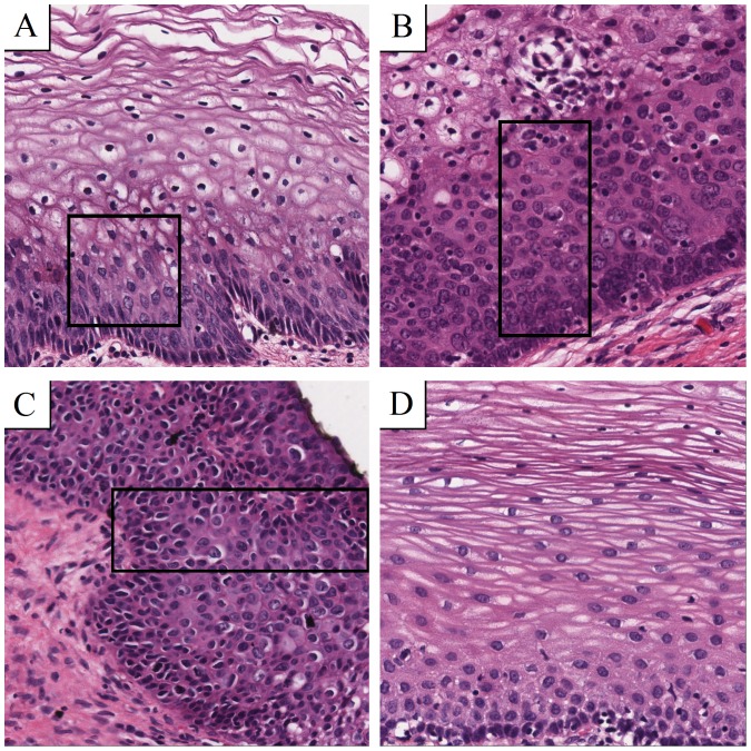
Figure 1. Morphology of CIN grades and normal epithelium.
All samples are Haematoxylin-Eosin (H&E) stained and digitally scanned at a magnification of 40. Boxes show witch area to focus on. (A) Atypical cells in the first third are typically for CIN 1. (B) CIN 2 contains atypical cells in the lower two thirds of the epithelium. (C) If the whole epithelium is covered by atypical cells, the grade is called CIN 3. (D) Atypical cells are missing in normal epithelium.

