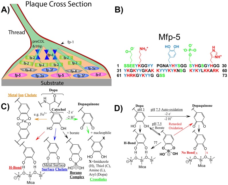
Figure 1. A schematic overview of Dopa-proteins and Dopa in mussel adhesion.
(A) Schematic cross-section of an adhesive plaque with mussel foot protein distribution. (B) Sequence of Mfp-5 highlighting distribution of Dopa (blue) and charged residues. (C) The versatile reactivity of Dopa in adhesion, cohesion and borate complexation. (D) The complexation of Dopa and borate to form an oxidation resistant Dopa-boronate species at pH 7.5 has critical implications for adhesion. Dopaquinone, which normally forms from the 2-electron oxidation of Dopa at pH 7.5 in the absence of borate, is unable to bond mica.

