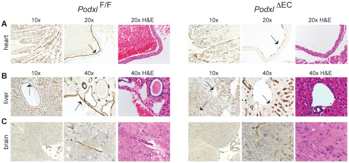
Figure 3. Cdh5-Cre fails to efficiently delete podocalyxin in a subset of organ vascular beds.
(A) In the wildtype heart podocalyxin is expressed by all endothelial cells. In the Podxl ΔEC mice, podocalyxin is efficiently deleted in large vessels such as the pulmonary artery (arrow, 20x mag. panels) but deletion in the smaller trabecular vessels of the heart muscle is variable (see 10x mag. panels). (B) In the liver of Podxl ΔEC mice, podocalyxin expression is ablated in the major vessels (portal and central veins, hepatic artery) but not in sinusoidal endothelial cells. (C) In the brain, podocalyxin is normally expressed in the ventricles and endothelial cells, including the microvasculature. Sections of the brain from Podxl ΔEC mice display similar staining to control mice indicating poor recombination of loxP sites by Cdh5-Cre in brain. Adjacent H&E sections demonstrate normal gross morphology podocalyxin-deficient tissues.

