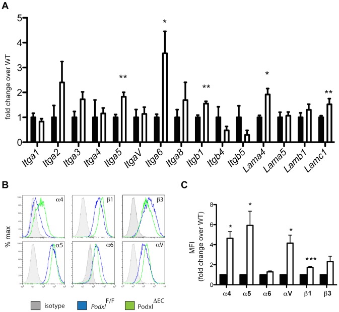
Figure 7. Podocalyxin deletion results in altered integrin and laminin expression in primary lung endothelial cells.
(A) Integrins gene expression in PodxlF/F (black bars) and Podxl ΔEC (white bars) cultured lung mEC (results are the mean of 3 independent mEC primary cultures per genotype). (B) Cell surface expression of integrins on lung mECs isolated from Podxl F/F (blue line) and Podxl ΔEC (green line) mice. Shown are representative histograms from flow cytometric assays from one experiment. (C) Surface expression levels of integrins were determined by flow cytometry using the mean fluorescence intensity (MFI) of the integrin staining in primary mEC cultures. The mean change in the MFI of Podxl ΔEC mECs (white bars) compared to Podxl F/F mEC (black bars, normalized to 1) are from 4 independently derived mEC cultures. Error bars = SEM, *Significantly different with P<0.05 or ***significantly different with P<0.005 using one-sample t test with hypothetical value set to 1 (normalized control).

