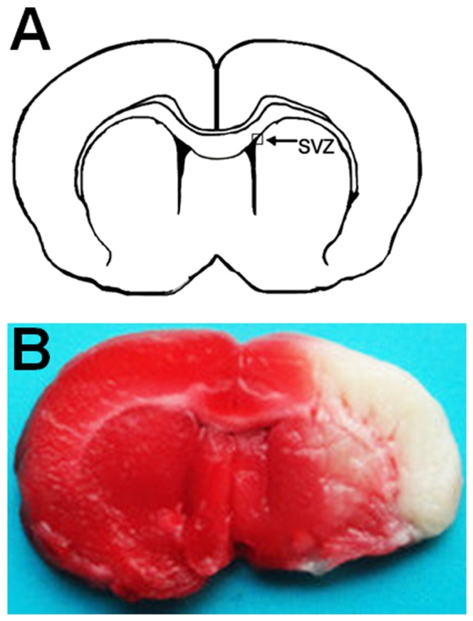
Figure 2. Infarct volume assessed by TTC staining 1 day after the tMCAO.

(A) Position of SVZ in the coronal section of brain. Areas imaged for immunofluorescence studies are indicated by box. (B) Coronal brain section stained by TTC 1 day after tMCAO. The white areas without deep red-staining indicate ischemic areas. SVZ, subventricular zone.
