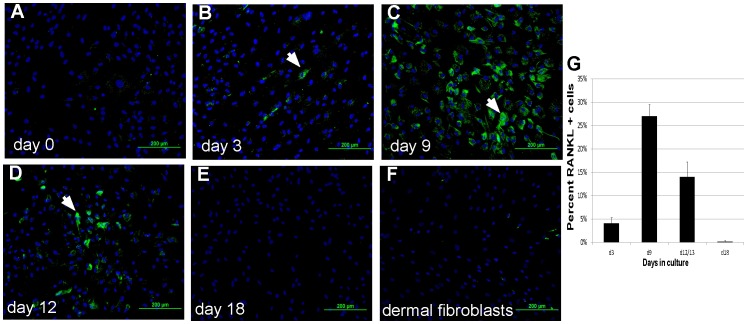
Figure 3. RANKL protein is regulated during adipocyte differentiation.
MSC cultures were induced to form MSC-derived adipocytes, and RANKL protein was detected using immunofluorescence. Cells were counterstained with Hoechst33342, and RANKL protein levels were quantified per cell number by manual counting. Analysis was done on (A) day 0, (B) day 3, (C) day 9, (D) day 12 and (E) day 18. (F) Dermal fibroblasts in culture between 3–12 days were used as a control. There is an increase in both the number and intensity (white arrows) of RANKL positive cells between days 3 and 12 after differentiation. Scale bar in panel A = 200 µm for all panels. (G) To quantify changes in RANKL protein levels cells, the percentage of stained cells was counted at different times in culture. n = 3–4 donors.

