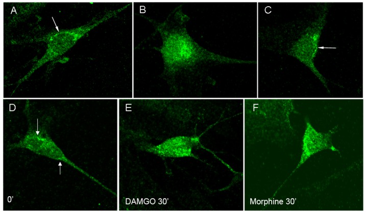
Figure 1. μOR immunoreactivity in naïve enteric neurons (A–C) and in neurons chronically treated with morphine (D–F).
μOR immunoreactivity is at the cell surface in unstimulated and morphine-stimulated neurons (A, C arrows), and it is in the cytosol following stimulation with DAMGO (B) in naïve enteric neurons. μOR immunoreactivity is at the cell surface in unstimulated neurons (D, arrows), but in the cytosol following DAMGO or morphine stimulation (E, F) in chronic enteric neurons.

