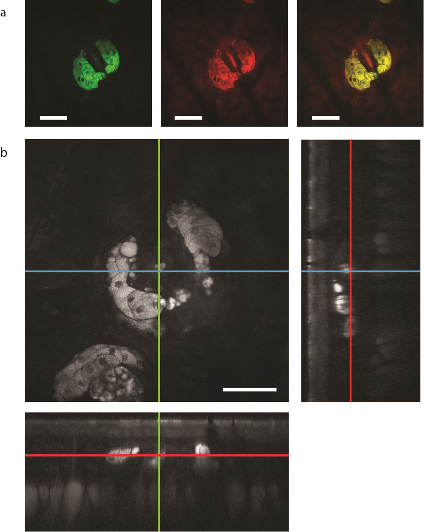
Figure 4. SRS spectral imaging with the fibre-laser source.
a, Two color image of a sebaceous gland in mouse skin acquired at 2850cm−1 (mainly lipids, green), 2950cm−1 (mainly proteins, red) and the composite of the two colours. b, z-stack acquired at 1 frame/s and cross sections from different directions. Scale bars, 50µm.

