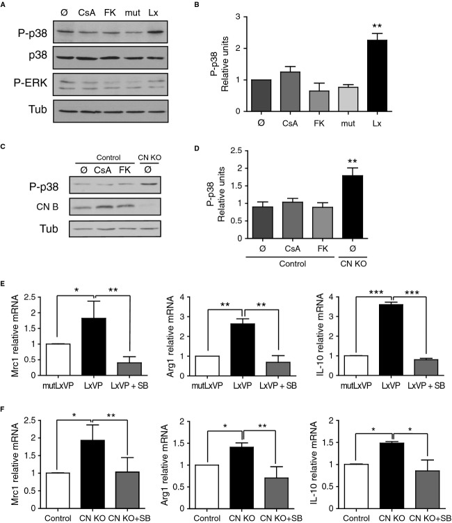
Figure 4. p38 MAPK activity mediates the induction of anti-inflammatory macrophages upon specific CN targeting.
A Representative Western blot showing expression of phosphorylated P-p38 and P-ERK in peritoneal macrophages treated with CsA or FK506 (FK) or transduced with LxVP or control lentivirus in vitro. p38 and tubulin were used as loading controls.
B Quantification of P-p38 expression in the experimental conditions mentioned in (A) (mean ± s.e.m.; n = 3).
C P-p38 (top) and CNB (center) protein expression in peritoneal macrophages from Cnb1 flox/flox LysMCre− (control) and Cnb1 Δ/flox LysMCre+ (CN KO) mice treated with CsA or FK506. Tubulin was used as a loading control (Tub).
D Quantification of P-p38 expression in the experimental conditions in (C) (mean ± s.e.m.; n = 3).
E, F Effect of p38 inhibition by SB203580 (SB) treatment on Mrc1, Arg1, and IL-10 mRNA levels in (E) LxVP-transduced macrophages and (F) CN KO macrophages.
Data information: *P < 0.05, **P < 0.01, ***P < 0.001.
Source data are available online for this figure.

