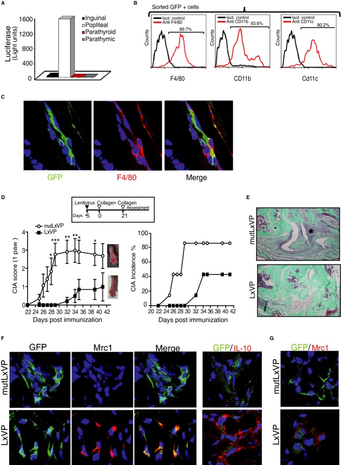
Figure 8. Locally delivered LxVP targets macrophages and prophylactically protects against CIA.
- Footpad-injected lentivirus targets popliteal lymph nodes (LN). The chart shows luciferase activity in different LN-cell homogenates from mice inoculated in the footpad with luciferase-encoding lentivirus.
- Flow cytometry analysis of F4/80, CD11b, and CD11c expression in sorted GFP+ cells obtained from popliteal LN after inoculation of footpads with GFP-encoding lentivirus.
- Confocal immunofluorescence showing colocalization of virus-encoded GFP (green) and F4/80 (red) in sections of inflamed paws locally injected with lentiviral vectors.
- The scheme shows the CIA protocol, indicating the time of lentivirus injection. Charts show time profiles of arthritic score (left) and incidence (right) in mice transduced in the right hind footpad on day −5 with LxVP or mutLxVP lentivirus. Data are means ± s.e.m. from a representative experiment of two; n = 10 mice (10 paws) per group.
- Masson's trichrome staining in joints of paws locally transduced on day −5 with mutLxVP or LxVP. Green: bone and collagen; blue: cell nuclei; red: muscle fibers and keratin. Asterisk marks inflammatory cell infiltrate.
- Confocal immunofluorescence showing GFP (green) and Mrc1 or IL-10 (red) in sections of arthritic paws locally injected with LxVP or mutLxVP lentivirus.
- Non-transduced cells express Mrc1 in LxVP-inoculated paws. Confocal immunofluorescence showing GFP (green) and Mrc1 (red) in arthritic paws locally injected with LxVP or mutLxVP lentivirus.
Data information: * P < 0.05; **P < 0.01; ***P < 0.001.

