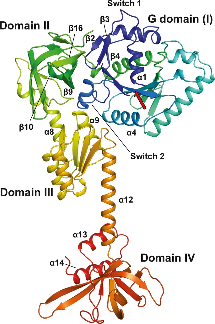
Figure 1. Front view of the overall structure of Chaetomium thermophilum eIF5B(517C) in the apo form.

The Cα trace is shown in rainbow coloring from the N- (blue) to the C-terminus (red). The functional core of eIF5B is composed of the G domain (I) with the nucleotide binding site (arrow), domain II, domain III and domain IV.
