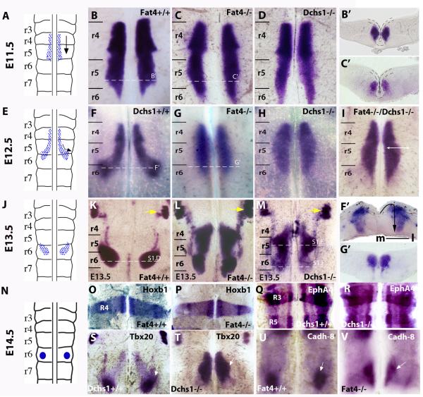
Figure 1. Fat4 and Dchs1 regulate lateral tangential FBM neuronal migration.
Sketch of FBM neuronal migration (A,E,J,N) and flat-mounts of a hindbrain whole-mounted for Islet1 (B-D, F-I, K-M), Hoxb1 (O,P), EphA4 (Q,R ), Tbx20 (S,T) and Cadherin 8 (U,V) expression in E10.5 (O-R), E11.5 (B-D), E12.5 (F-I, S-V), or E13.5 (K-M) wildtype (B,F,K,O,Q,S,U), Fat4−/− (C,G,L,P,V), Dchs1−/− (D,H,M,R,T) or Fat4−/− Dchs1−/− (I), embryos. The trigeminal nucleus is indicated by a yellow arrow in (K-M). The dashed lines indicate levels of sections shown in B’,C’,F’,G’ and in Fig. S1D-F; the ventricular layer is uppermost, the midline is outlined in B’,C’. The arrow in F’ indicates the direction of FBM neuronal migration: initially there is a tangential lateral migration followed by a radial migration from the ventricular layer to the pial surface. (B-D) and (F-H) are viewed from the ventricular surface; and (K-M) from the pial surface. r3-r7, rhombomeres 3-7. The FBM neurons are arrowed in S-V; Cadh-8 is expressed at the leading edge of the FBM neurons in wildtype (U) and Fat4−/− (V) embryos. m, medial; l, lateral

