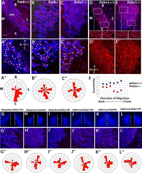
Figure 2. FBM neurons fail to polarise in Fat4−/− and Dchs1−/− mutants.
(A-C) Immunolocalisation showing the polarity of the Golgi (red) in migrating FBM neurons (blue, Islet1 staining) at E12.5 in wildtype (A), Fat4−/− (B) and Dchs1−/− (C) embryos. A’-C’ are higher power images of the leading (caudal) edge. The arrows and arrowheads indicate the long axis of the nucleus and point towards the position of the Golgi apparatus. The angles of the arrows relative to the midline were scored for each neuron and quantified in the Rose Plots below each image (A”-C”). In wildtype embryos the FBM neurons become polarised caudal-laterally at E12.5. This polarisation does not occur in Fat4−/− or Dchs1−/− mutants. (D, E) Phalloidin staining (red) of the FBM neurons (blue, Islet1 staining) in wildtype (D) and Dchs1−/− (E) E12.5 embryos. Outlines of the cell shapes at the leading edge and FBM neurons in the centre of the neuronal stream are shown in the insets below. The cell shape of the FBM neurons is quantified in (F) where 0 is a circle and 1 is an ellipse. D’, E’ show the phalloidin staining alone. Wildtype FBM neurons display actin cables aligned in the direction of migration while Dchs1−/− FBM neurons show a less organised F-actin cytoskeleton. (G-L) Tissue-specific requirements of Fat4 and Dchs1 within the neuroepithelium and the FBM neurons. Low power images of FBM neuronal migration (Islet1 staining, blue) at E12.5 (G-L) together with higher power images of the leading caudal edge of the FBM neuronal stream showing Islet1 (blue) and Golgi (red) immunostaining (G’-L’) and Rose plots of Golgi orientation (G”-L”), in Hoxa3CreDchsf/+ (G), Hoxa3CreFat4f/− (H), Hoxa3CreDchsf/− (I), Islet1CreDchs1f/+ (J), Islet1CreFat4f/− (K) and Islet1CreDchs1f/− (L) E12.5 embryos. The midline is indicated by the two white dashed lines in (G-L). R, rostral; C, Caudal; M, medial; L, Lateral.

