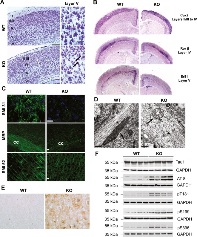
Figure 3. Constitutive Afg3l2 deletion leads to microtubule fragmentation and tau hyperphosphorylation.
- Nissl staining of coronal sections across the cerebral cortex of Afg3l2 knockout (KO) and wild-type (WT) show normal lamination but degenerating neurons in layer V (arrows).
- In situ hybridization of brain coronal sections of WT and KO using the indicated probes.
- Immunofluorescence analysis of coronal sections across the cerebral cortex of Afg3l2 KO and WT mice with the indicated antibodies shows dramatic loss of myelinated axons. cc: corpus callosum.
- Electron micrographs of cortical neuronal processes show microtubule fragmentation and disorganization, and abnormal mitochondria in KO mice. Arrows: mitochondria; asterisks: microtubules.
- Cerebral cortex sections were stained with AT8 antibody. Phosphorylated tau accumulates in cell bodies and dendrites of KO mice.
- Western blot analysis of brain lysates from Afg3l2 KO and WT littermates using the indicated antibodies. KO brains display increased levels of phosphorylated tau species.
Data information: Scale bar in (A) 200 μm, in (C) 20 μm, in (D) 0.5 μm.
Source data are available online for this figure.

