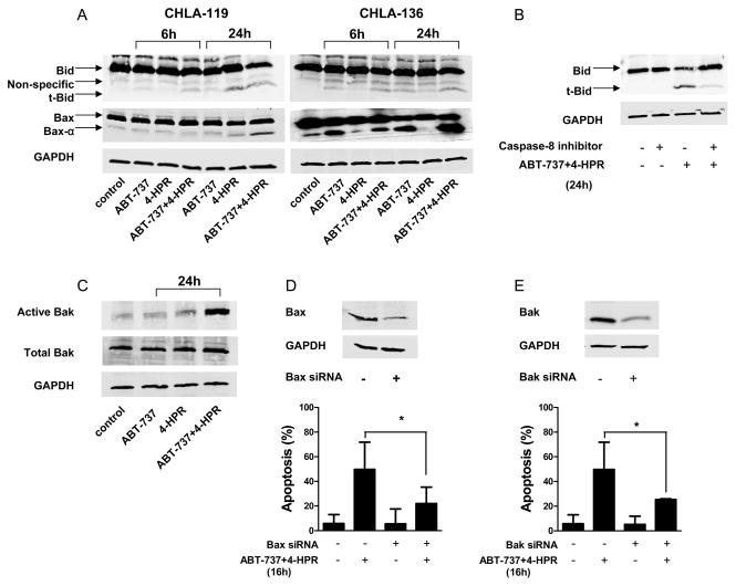
Figure 4. Effects of ABT-737 plus 4-HPR on the activation of pro-apoptotic proteins.
CHLA-119 or CHLA-136 cells were incubated with ABT-737 or 4-HPR (2.5, 5 μM, respectively) or the combination for 6 or 24 hours. (A) Cell lysates were then examined for the presence of pro-apoptotic Bid and Bax. (B) CHLA-119 cells were pretreated with Z-IETD-FMK (20 μM) for 1 hour before being exposed to ABT-737+4-HPR (2.5 μM each) for 24 hours. Cell lysates were then examined for Bid cleavage into t-Bid by western blot analysis. (C) CHLA-119 cells were incubated with ABT-737 or 4-HPR (2.5 μM each) or the combination for 24 hours, and the presence of conformationally active Bak and total Bak were detected by western blot. (D and E) CHLA-119 cells were transfected with siRNA against Bax or Bak or control siRNA as indicated in materials and methods. Knockdown of Bax (D) and Bak (E) protein expression was assessed by western blot (upper panels). 48 hours after transfection, cells were treated with ABT-737+ 4-HPR (2.5 μM each) for another 16 hours and apoptosis was determined by flow cytometric TUNEL assay (lower panels). The bar graph shows the mean percentage of apoptotic cells (± SD) of triplicate samples. All the data shown are representative of two independent experiments showing similar results. * P < 0.05.

