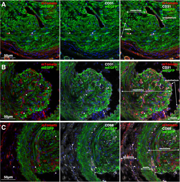
Figure 9.
CD31 and CD68 positive cells in mature neointimal lesions. Images shown are examples of mature lesions seen in 2 different Myh11 creER(T2)-/+ mTmG-/+ mice 28 days following ligation (Mouse 4(A) and 6(B, C); Table 1). A, B. Sections were immuno-stained for the endothelial/platelet marker CD31. Images shown are merged images showing the mTomato (red), mEGFP (green) and DAPI (blue); CD31 (white), mEGFP (green) and DAPI (blue) or mTomato, mEGFP, CD31 and DAPI channels, as indicated. Sections obtained from the mouse shown in panel A exhibited very few CD31 positive cells in the mature neointima; in contrast the lesion shown in panel B exhibited numerous CD31 positive, mTomato positive, mEGFP negative cells in the neointima (examples indicated by arrow heads). C. Sections (serial to those shown in panel B) were immuno-stained for the macrophage/monocyte marker CD68. Images shown are merged images showing the mTomato (red), mEGFP (green) and DAPI (blue); CD68 (white), mEGFP (green) and DAPI (blue) or mTomato, mEGFP, CD68 and DAPI channels, as indicated. Several CD68 positive, mTomato positive, mEGFP negative cells can be seen in the neointima and adventitia (examples indicated by arrow heads). No CD68 positive cells were seen in the neointima of the mouse shown in panel A (data not shown).

