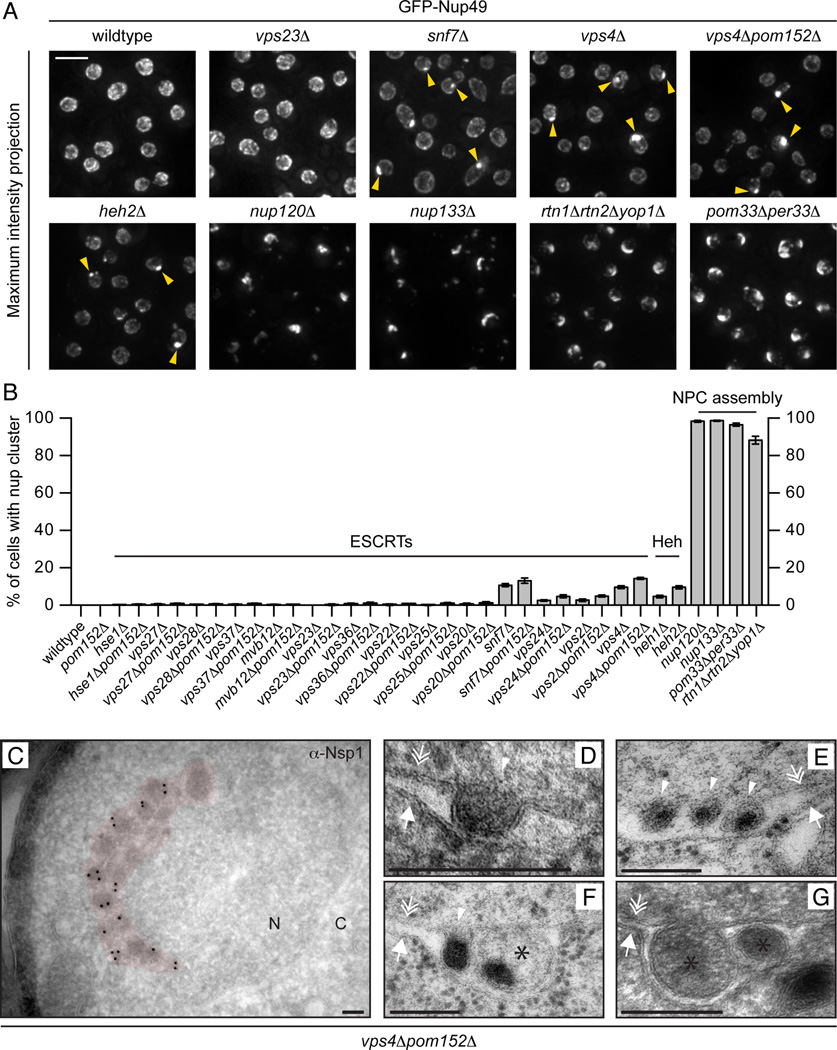
Figure 4. Nups accumulate in a cluster at the NE in the absence of ESCRT-III/Vps4.
(A) GFP-Nup49 accumulates at one side of the NE in snf7Δ and vps4Δ strains. Maximum intensity projections shown. Arrowheads point to the nup cluster. Bar is 5µm.
(B) Plot of the percentage of cells in the indicated strains where GFP-Nup49 accumulates in a cluster. Mean +/− s.d. from 3 experiments (n>400).
(C-G) TEM micrographs of vps4Δpom152Δ cells showing an accumulation of NPC-like structures on one side of the NE (pseudo-colored red) in addition to intralumenal vesicles. All bars are 200 nm. In (C), an anti-Nsp1 antibody and 10nm-gold conjugated secondary antibodies label the NPC-like structures. “N” and “C” represent nucleoplasm and cytoplasm, respectively. Arrows, double arrows and arrowhead point to the ONM, INM and INM invaginations, respectively. Asterisks label putative vesicles in the NE lumen.

