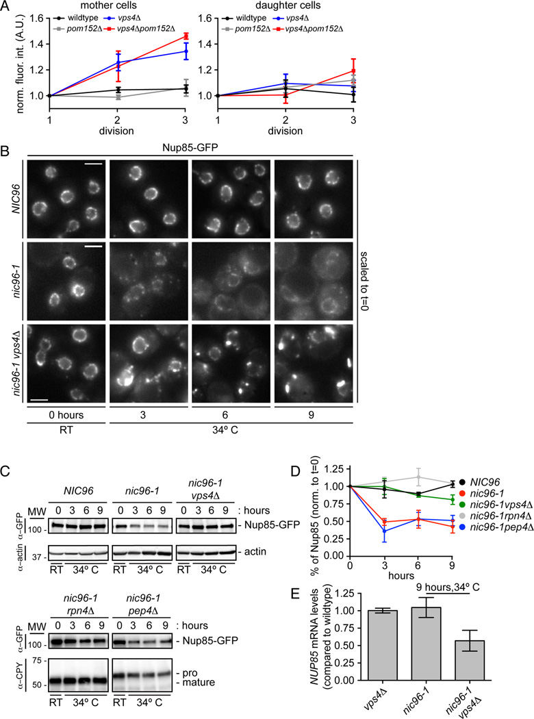
Figure 7. Vps4 destabilizes misassembled nups.
(A) An extra pool of Nup85 accumulates in the SINC of vps4Δ mothers. Plot of total Nup85-GFP fluorescence in mother (left) and daughter (right) nuclei measured after each of three divisions (normalized to first). Mean +/− s.d. from 3 experiments in which 5 cells (mother and daughter) were measured.
(B) Nup85-GFP is stabilized in vps4Δ cells when NPC assembly is inhibited. Micrographs of Nup85-GFP at either RT or at 34°C. Bars are 5µm.
(C) Nup85-GFP is degraded by the proteasome when NPC assembly is inhibited. Western blot of Nup85-GFP levels (with actin or CPY load controls) from experiments identical to that shown in B.
(D) Plot of the quantitation of Nup85-GFP levels in the indicated strains. Mean +/− s.d. from 3 experiments.
(E) NUP85 transcript levels decline in nic96-1vps4Δ cells. RT-qPCR analysis of NUP85 transcript compared to WT cells.
See Figure S5.

