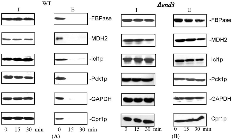
Figure 4.
The decline of extracellular FBPase, MDH2, Icl1p, Pck1p, GAPDH, and Cpr1p in response to glucose re-feeding is dependent on END3. Wild-type cells (A) and the ∆end3 cells (B) were starved of glucose for 3 days and re-fed with glucose for 0, 15, and 30 min. Western blotting was used to examine levels of FBPase, MDH2, Icl1p, Pck1p, GAPDH, and Cpr1p in the intracellular (I) and extracellular (E) fractions. This figure is in a manuscript published in the Journal of Extracellular Vesicles [93].

