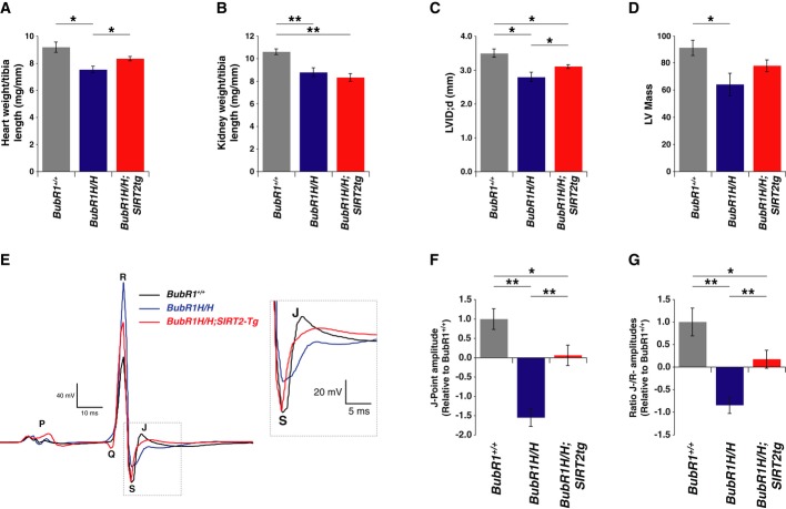
Figure 5. Reversal of cardiac defects in BubR1H/H mice by SIRT2.
- Hearts from male 3-month-old wild-type (n = 6), BubR1H/H (n = 5), and SIRT2tg/BubR1H/H (n = 6) mice were weighed and normalized to tibia length.
- Kidneys analyzed as in (A).
- LVID;d measured by echocardiography.
- Left ventricle mass calculated with echocardiography measurements.
- Representative ECG tracings from 3-month-old wild-type, BubR1H/H, and SIRT2tg/BubR1H/H mice with inset highlighting J-point. Electrocardiography measurements from male 3-month-old wild-type (n = 12), BubR1H/H (n = 14), and SIRT2tg/BubR1H/H (n = 12) mice representing (F) J-point amplitudes and (G) normalization of J-point to R-wave amplitude.
Data information: Error bars represent SEM. P-values calculated using Student’s t-test (n as specified), *P < 0.05, **P < 0.005.

