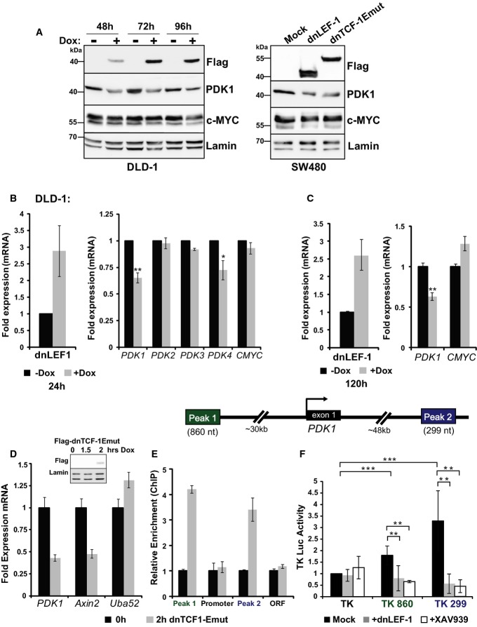
Figure 4. Blocking Wnt directly reduces PDK1 levels via regulation of transcription.
A Whole-cell lysates from DLD-1 dnLEF-1(1) cells were collected 48, 72, and 96 h after 0.01 μg/ml doxycycline treatment and were probed with the antibodies shown. SW480 cells were harvested 48 h post-transduction.
B, C 9RT-qPCR analysis was performed on RNA collected from DLD-1 dnLEF-1(2) cells harvested 24 h (B) or 120 h (C) after the addition of doxycycline. Graphs shown represent the average of three trials (± SEM).
D RT-qPCR analysis was performed on 4-thiouridine-labeled RNA isolated from a 30-min pulse in the presence/absence of dnTCF-1Emut, induced by 2-h doxycycline treatment in DLD-1 cells. Known Wnt target genes Axin2 and Uba52 were used as positive and negative controls, respectively. A representative graph is shown of two replicates, with error bars representing the SD among three internal replicates.
E RT-qPCR analysis of chromatin immunoprecipitated from DLD-1 cells with or without induction of FLAG-dnTCF-1Emut, using anti-FLAG antibody. PCR primers designed to detect the indicated PDK1 genomic regions show that dnTCF-1Emut associates with distal regions that flank the PDK1 locus. A representative graph is shown of two replicates, with error bars representing the SD among three internal replicates.
F Luciferase reporter activity in SW480 cells shows that Peak 1 and Peak 2 regions confer elevated transcription activity to the heterologous thymidine kinase (TK) promoter. Expression of transduced dnLEF-1, or treatment with the Wnt inhibitor XAV939 (10 μM), eliminates the regulatory activity of these fragments. Graph shown represents the average of three independent replicates (± SD).
Data information: *P-value < 0.05; **P-value < 0.01; ***P-value < 0.001).
Source data are available online for this figure.

