
Figure 1. Src upregulation drives ISC proliferation.
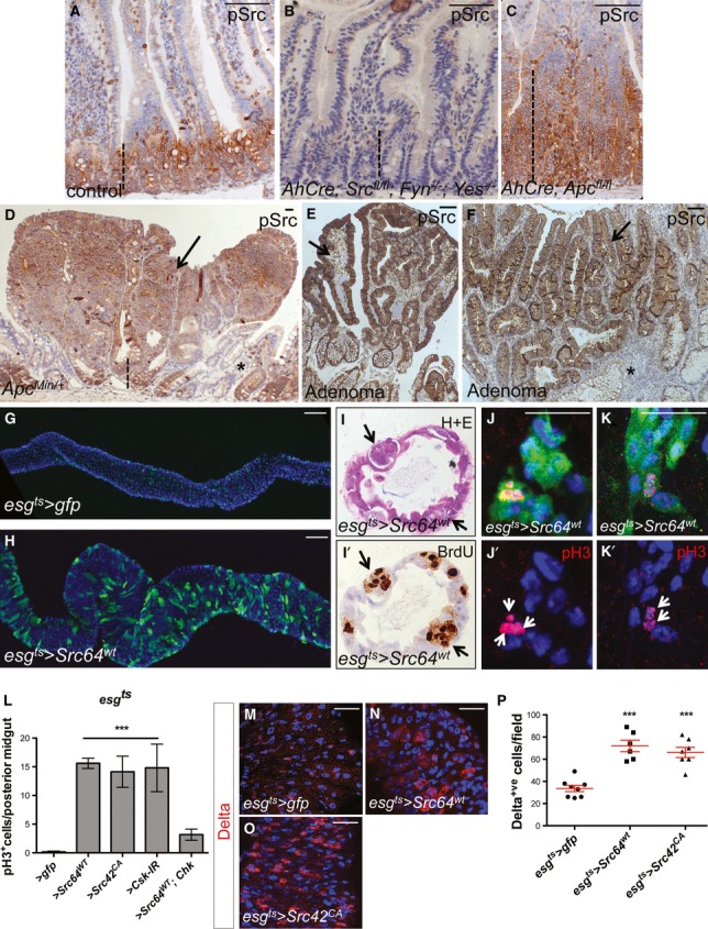
A–D Immunohistochemistry to detect the activated form of Src (pSrc) in tissue sections from mouse and human intestines. pSrc is detected in the proliferative ‘crypt’ region (indicated with dashed line) at the base of the mouse small intestinal epithelium in control animals (A). Conditional knockout of Src (Srcfl/fl) combined with full knockout of related kinases Fyn and Yes, resulted in no staining within the intestinal epithelium (B). Intestines with conditional Apc knockout (Apcfl/fl) depict the expected ‘crypt-like’ progenitor phenotype (dashed line) and expansion of the pSrc domain (C). Example of a small intestinal polyp from an ApcMin/+ mouse, showing high p-Src staining within the core of the polyp (arrow), is shown in (D). Note normal tissue around polyp showing pSrc localized at the crypt base (dashed line). Scale bars, 100 μm.
E, F pSrc is upregulated within benign human intestinal adenoma lesions (arrows) when compared with normal surrounding tissue (asterisks). Scale bars, 100 μm.
G, H Adult Drosophila midguts overexpressing gfp (G; control) or Src (H) for 7 days under the stem/progenitor cell (ISCs/EBs) driver escargot-gal4 (esgts > gfp and esgts > Src64wt, respectively). Unless otherwise noted green marks esg > gfp cells and Dapi (blue) stains all cell nuclei. Scale bars, 100 μm.
I, I’ Paraffin-embedded sections from 7-day-old Src-overexpressing midguts (esgts > Src64wt) analysed by Haematoxylin and Eosin H+E (I) and BrdU (I’) staining. Arrows point to ‘polyp-like’ structures containing BrdU+ve cells.
J–K’ 7-day-old esgts > Src64wt posterior midguts stained with anti-pH3 (red) to visualize proliferating ISCs (arrows). Scale bars, 20 μm.
L ISC proliferation quantified as the number of cells which stained positive for phosphorylated histone 3 (pH3) in posterior midguts from control animals (esgts > gfp) or animals overexpressing Src (esgts > Scr64wt or esgts > Src42CA) or RNA interference for the Src inhibitor Csk (esgts > Csk-IR). Note that co-expression of human ChK (esgts > Scr64wt; Chk) suppresses Src-driven hyperproliferation in the Drosophila midgut.
M–O 7-day-old adult posterior midguts from animals of the indicated genotypes stained with anti-Delta (red) to label ISCs. Scale bars, 20 μm.
P Quantification of the number of Delta+ve ISCs per field from posterior midguts as in (M-O).
Data information: Data in (L) and (P) represent average values ± SEM (***P < 0.0001 one-way ANOVA with Bonferroni’s multiple comparison test)