
Figure 6. Activation of cellular signalling with spatial precision.
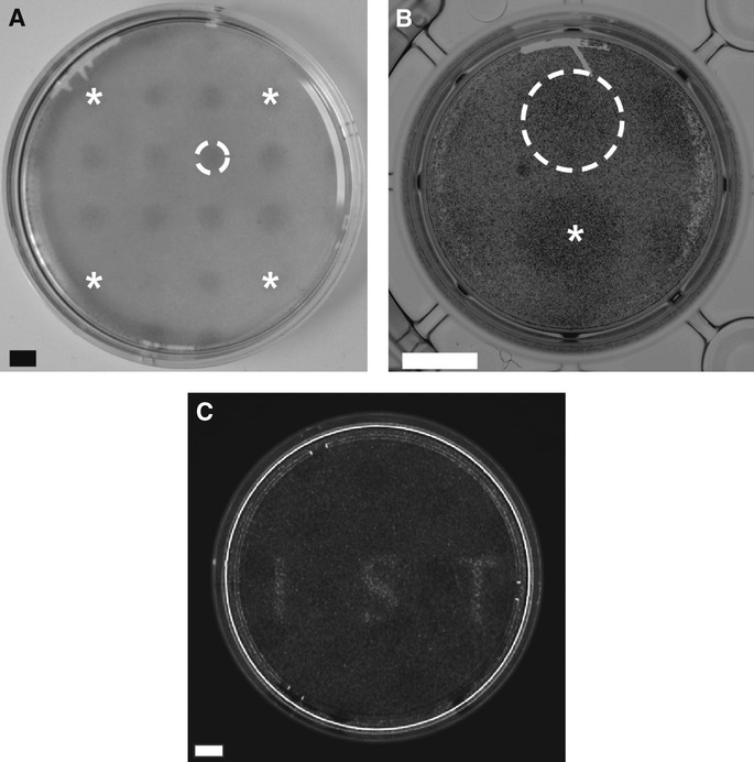
- Spatially confined ERK1/2 phosphorylation in SPC212Opto-mFGFR1 cells. A illumination pattern of 16 equidistant circles was produced using a pinhole template. Stars mark circles in corners. Dashed line marks a single circle. Cells were stimulated with blue light for 5 min followed by fixation and staining. Scale bar: 2 mm.
- Spatially confined ERK1/2 phosphorylation in hBEOpto-mFGFR1 cells. A illumination pattern of two circles was produced using a pinhole template. Star marks the center of one circle, dashed line marks the other circle. Cells were stimulated with blue light for 5 min followed by fixation and staining. Scale bar: 5 mm.
- Spatially confined MAPK-dependent gene transcription in HEK293 cells. Thirty-four hours after transfection, cells stimulated with light for 8 h followed by live-cell detection of luciferase. Scale bar: 10 mm.
Data information: All images are unprocessed raw images. Light intensity was ˜2.5 μW/mm2.