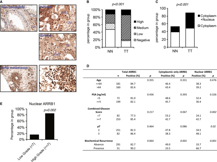
Figure 1. Nuclear ARRB1 is increased in prostate cancer.
- Representative expression pattern of ARRB1 in non-neoplastic and malignant prostate cancer tissues. Non-neoplastic tissue shows weak nuclear and moderate cytoplasmic staining in luminal and basal cells. Staining is also present in stromal cells (s). Moderate to intense cytoplasmic and intense nuclear staining is noted in Gleason 4 (G4) areas of the tumour. Intense staining is noted in scattered bone metastatic prostate cancer cells.
- Quantification of ARRB1 staining in non-neoplastic and malignant prostate tissue shown in (A) (Porto TMA, see Supplementary information for details). NN=non-neoplastic, TT=tumour tissue. P < 0.001 for total positive ARRB1 cases in TT versus NN.
- Nuclear (solely nuclear + cytoplasmic and nuclear) or solely cytoplasmic ARRB1 staining in non-neoplastic and tumour tissue. P < 0.001 for positive nuclear ARRB1 in TT versus NN.
- Assessment of association between ARRB1 expressions (total expression versus only cytoplasmic versus nuclear) in prostate cancer samples and clinicopathological data. The comparisons were examined for statistical significance using Pearson's chi-square (χ2) test, P < 0.05 being the threshold for significance.
- Distribution of nuclear ARRB1 in low (< 7) and high (≥ 7) grade tumours.

