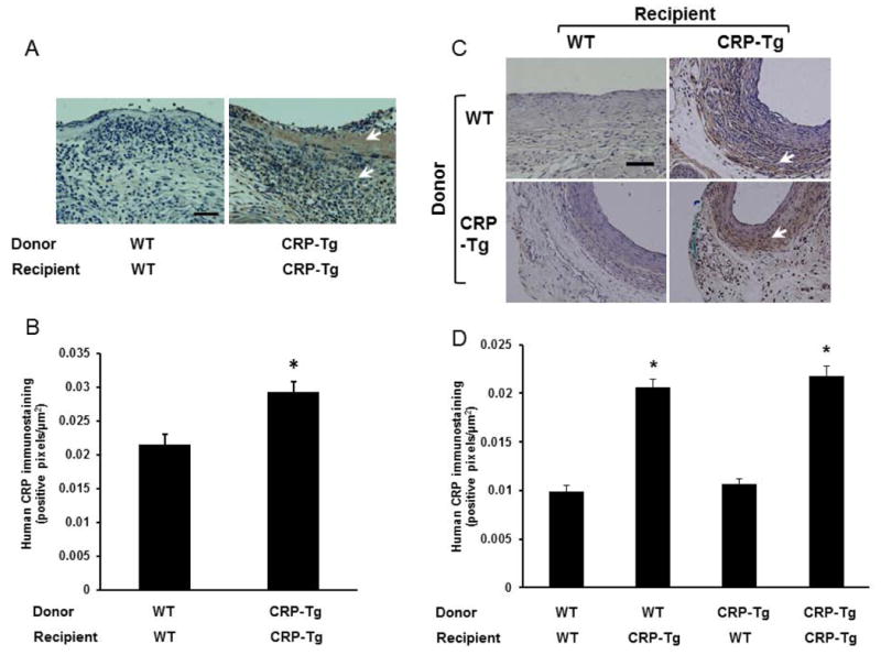
Figure 2.

Human CRP immunostaining in VGs. (A) VGs harvested 7 days after surgery. Donor/recipient genotypes are as shown. (B) Quantitative analysis of intimal CRP immunostaining at 7 days after surgery (n=4 mice/group). *P<0.02 vs. WTdonor/WTrecipient group. (C) VGs harvested 28 days after surgery. For this time point all 4 potential combinations of donors and recipients were included. (D) Quantitative analysis of intimal CRP immunostaining at 28 days after surgery (n=3 mice/group). *P<0.01 vs. WTdonor/WTrecipient group. In panels (A) and (C) brown color (arrow heads) represents CRP immunostaining; scale bars = 50 μm.
