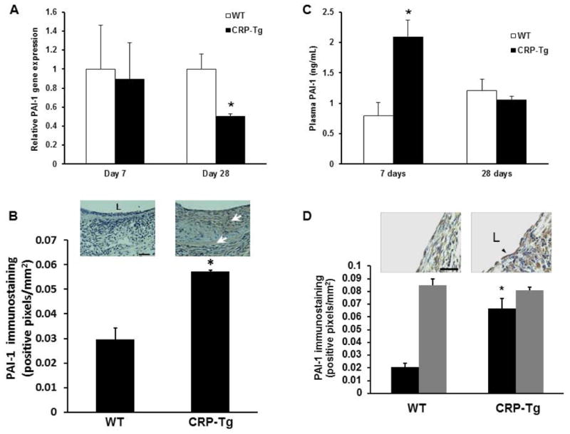
Figure 5.

Effects of human CRP on PAI-1 expression. (A) Human CRP does not increase total PAI-1 gene expression in VGs (on Day 7, P>0.8; n=6/group; on Day 28, PAI-1 gene expression is significantly less in VGs of CRP-Tg mice than WT controls, *P<0.05, n=4/group). (B) Immunohistochemical analysis of PAI-1 protein in VGs 7 days after surgery. PAI-1 immunostaining (brown color, arrows) is significantly greater in VGs of CRP-Tg mice compared to VGs of WT mice (*P<0.02, n=4/group). Representative images are shown. Distance bar = 50 μm. “L” denotes lumen. (C) Plasma PAI-1 concentration is higher in CRP-Tg mice than in WT mice on day 7 after surgery, but not on day 28. *P<0.01 vs. WT mice; n=6/group at 7 days; n=4/group at 28 days. (D) On day 28 after surgery PAI-1 immunostaining is significantly greater in endothelial cells (black bars) of CRP-Tg VGs compared to WT controls (*P<0.01 vs. WT mice, n=4/group), whereas PAI-1 immunostaining within the subendothelial intima (gray bars) does not differ significantly between groups. Representative images are shown. Arrow head points to endothelial cells. Scale bar = 25 μm.
