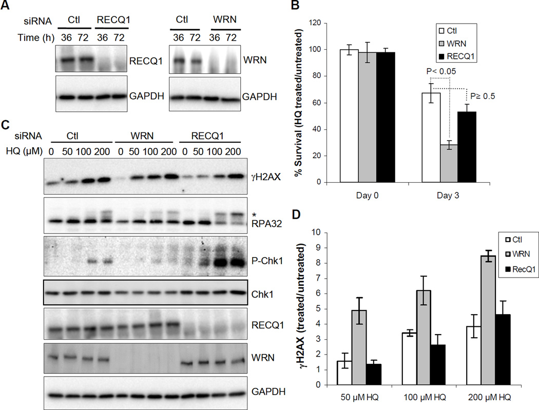
Figure 1. Cell survival and response to HQ treatment.
A. Depletion of RECQ1 and WRN in HeLa cells. Western blot shows efficient knockdown of WRN or RECQ1. B. Cell survival following HQ treatment (50 µM, 72 h). Surviving fraction is presented as the mean ± SD of three independent experiments. Statistical significance of differences in survival is indicated by P value. C. DNA damage response. Cells were treated with indicated dose of HQ for 24 h and equal amounts of total protein were used for Western blotting with indicated antibodies. Phosphorylated RPA32 is indicated by asterisk. D. HQ-induced γH2AX levels. Signals intensities, quantitated using ImageJ, were normalized with GAPDH and the fold enrichment in γH2AX was calculated. Mean ± S.E.M. from three independent experiments are shown. Ctl, control; pChk1, phospho-Chk1(Ser345); GAPDH serves as loading control.

