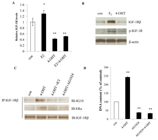
Figure 5. 4-OHT increased phosphorylation of IGF-1R in MCF-7:PF cells.
(A) Expression of IGF-1Rβ mRNA. MCF-7:PF cells were treated with vehicle (0.1% EtOH), E2 (10−9 mol/L), 4-OHT (10−6 mol/L), or E2 (10−9 mol/L) plus 4-OHT (10−6 mol/L) for 72 hours. IGF-1Rβ mRNA was quantitated through real-time RT-PCR. p<0.05, * compared with control, p<0.001, ** compared with control. (B) Protein levels of p-IGF-1Rβ and total IGF-1Rβ. MCF-7:PF cells were treated with vehicle (0.1% EtOH), E2 (10−9 mol/L), or 4-OHT (10−6 mol/L) for 72 hours. Total IGF-1Rβ and p-IGF-1Rβ were determined by Western blot. (C) Interaction between ERα and p-IGF-1Rβ. MCF-7:PF cells were treated with vehicle (0.1% EtOH), 4-OHT (10−6 mol/L), 4-OHT (10−6 mol/L) plus ICI (10−6 mol/L), and 4-OHT (10−6 mol/L) plus AG1024 (10−5 mol/L) for 24 hours. Protein-protein interaction was determined by immunoprecipitation. (D) Growth response to the IGF-1R inhibitor. MCF-7:PF cells were treated with vehicle control (0.1% DMSO), 4-OHT (10−6 mol/L), AG1024 (10−5 mol/L), and AG1024 (10−5 mol/L) plus 4-OHT (10−6 mol/L) for 7 days. Total DNA was determined as above. p<0.001, ** compared with control.

