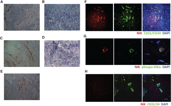
Figure 2.
NIK is expressed in endothelial cells in tumour tissues. NIK expression in (A) renal carcinoma tissue, (B) breast cancer tissue, (C) pancreatic cancer tissue, (D) melanoma tissue, (E) colorectal cancer tissue, and (F) IF staining on NIK (red), CD31/CD34 (green), and nuclei (blue) in breast cancer tissue. (G) IF staining on NIK (red), phospho-IKKα (green), and nuclei (blue) in breast cancer tissue. (H) IF on NIK (red), CD31/CD34 (green), and nuclei (blue) in normal skin. Representative pictures are shown (n = 5–6 patients or healthy donors, two independent experiments). Scale bars: (A–F, H) 100 µm; (G) 11.2 µm.

