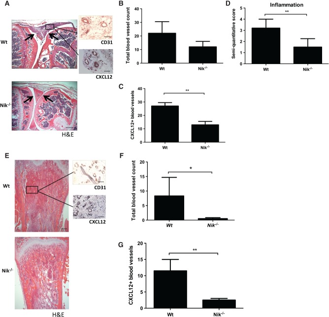
Figure 5.
Nik−/− mice exhibit reduced inflammation-induced angiogenesis in arthritis and reduced tumour-associated angiogenesis in a melanoma model. (A) Representative picture of an mBSA-injected Wt mouse knee joint showing a severe inflammatory infiltrate (between arrows), in contrast to an mBSA-injected Nik−/− knee joint showing a reduced inflammatory infiltrate and intact cartilage (between arrows: scale bar = 500 µm). Sagittal sections of knee joint with H&E staining and CD31 or CXCL12 IHC staining (insets: scale bars = 100 µm). (B) Quantification of the total number of synovial blood vessels in Wt and Nik−/− mice. (C) Quantification of CXCL12+ blood vessels in synovial tissue of Wt and Nik−/− mice. (D) Quantification of synovial inflammation on a semi-quantitative scale of 0–4. (E) Representative pictures of tibial sections from Wt and Nik−/− mice, with B16 melanoma tumour tissue in trabecular bone. H&E-stained sections of tibias show marrow replacement by tumour cells beneath the growth plate (scale bar = 500 µm), and insets show CD31 and CXCL12 IHC staining on tumour tissue (scale bar in insets = 100 µm). (F) Quantification of the total number of blood vessels in melanoma tumour tissue in Wt and Nik−/− mice. (G) Quantification of CXCL12+ blood vessels in melanoma tumour tissue of Wt and Nik−/− mice. (A–D) n = 5 per group; (E–G) n = 6 per group. (B–D, F, G) Mean ± SEM. *p < 0.05; **p < 0.001.

