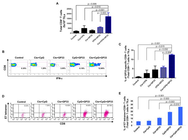
Figure 4. Local immune response to GP33 peptide and tumor antigen (HPV E7).
C57BL/6 mice (3 mice/group) were challenged subcutaneously with TC-1 tumor cells and treated with various combinations of cisplatin, CpG and GP33 peptide as indicated. 12 days after the last antigen delivery, tumor-infiltrating lymphocytes were harvested to analyze the immune cell subsets. A. Representative flow cytometry analysis depicting the frequency of GP33-specific IFN-γ-secreting CD8+ T cells among tumor infiltrating CD8+ T cells. B. Representative flow cytometry analysis depicting the absolute number of E7-tetramer binding CD8+ T cells in 5×10⁵ single-cell suspension prepared from tumors. C. Bar graph quantification of the absolute number of total CD8+ T cells in 5×10⁵ single-cell suspension prepared from tumors (mean ± S.E.). D. Bar graph quantification of the percentage of GP33-specific IFN-γ-secreting CD8+ T cells among tumor infiltrating CD8+ T cells (mean ± S.E.). E. Bar graph depicting the number of H2-Db tetramer+ CD8+ T cells in 5×10⁵ tumor infiltrating cells (mean ± S.E.).

