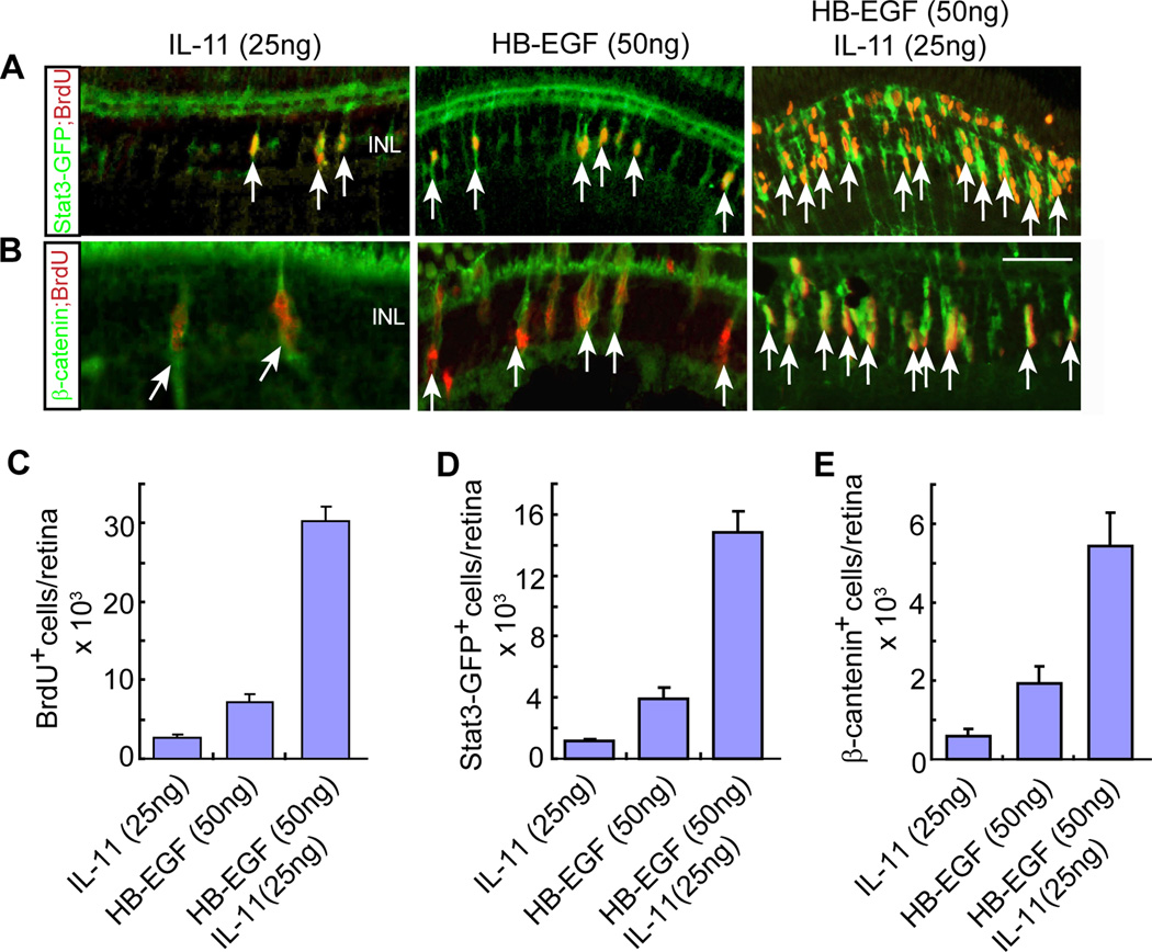
Figure 5. HB-EGF and IL-11 synergize with each other to stimulate MG proliferation and activation of the Stat3 and β-catenin signaling components in the uninjured retina.
(A, B) HB-EGF and IL-11 synergize with each other to stimulate the generation of BrdU+ progenitors and the accumulation of Stat3 (A) and β-catenin (B) signaling components in the uninjured retina of gfap:stat3-gfp transgenic fish. Arrows point to double-labeled cells. Scale bar is 50 µm. The green immunofluorescence signal in the photoreceptor layer is non-specific since it does not show up when using a secondary antibody coupled to a red Fluor (Figure S7). (C–E) Quantification of BrdU+ (C), Stat3-GFP+ (D) and β-catenin+ (E) progenitors in retinas intravitreally injected with HB-EGF, IL-11 or HB-EGF/IL-11. Error bars are SD; n=3. See also Figure S5.

