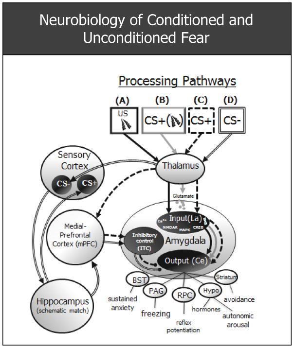
Figure 1.
Schematic of fear circuitry subserving unconditioned and conditioned fear derived from animal data. (A) Solid black arrows mark the processing path for an inherently aversive unconditioned stimulus (US: e.g., electric shock) which activates the input and output nodes of the amygdala, culminating in the behavioral, endocrine, and autonomic constituents of the fear response. (B) The light grey processing path subserves acquisition of conditioned fear, ensuing when a neutral conditioned stimulus (CS+) causes the release of glutamate in LA neurons in close temporal proximity to US-induced depolarization of these same neurons. The co-occurrence of this glutamate release and depolarization results in a cascade of intracellular processes in the La (release of Ca2+ which binds to NMDA receptors → excitation of protein kinases [e.g., MAPK] → activation of transcription factors [CREB] → synthesis of RNA and protein) which then makes new proteins used, in part, to increase the amygdala’s sensitivity to the CS+. (C) Broken arrows delineate extinction of conditioned fear, whereby repeated presentations of the CS+ in the absence of the US weakens the conditioned fear response. This weakening is thought to ensue when an mPFC-ITC mediated inhibition of the central nucleus of the amygdala outcompetes the excitatory pathway formed during acquisition. (D) Denoted by compound arrows is the processing path for discrimination of conditioned fear to a conditioned safety cue (CS-) with perceptual resemblance to the CS+. Sensory and affective discrimination of CS− from CS+ begins with higher order processing of CS− in the sensory cortex. Next the hippocampus may perform a schematic match between the cortical representation of the previously encountered CS+ and that of the currently presented CS−. With sufficient mismatch, the hippocampus initiates pattern separation resulting in activation of the mPFC, which then inhibits fear by activating a group of inhibitory, intercalated (ITC) neurons in the amygdala. La = lateral nucleus of the amygdala; Ce = central nucleus of the amygdala; Ca2+ = calcium; NMDAR = N-Methyl-D-aspartic acid receptor; MAPK = Mitogen-activated protein kinase; CREB = cAMP response element-binding transcription factor; BST = bed nucleus of the stria terminalis; PAG = periaqueductal gray; RPC = reticularis pontis caudalis; hypo = hypothalamus;
 = excitatory connections;
 = inhibitory connections.

