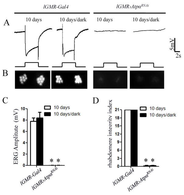
Figure 5.
Photoreceptor degeneration and the loss of light response in 10-day-old ATPα knockdown flies are independent of light exposure. A, No ERG difference was observed between light-exposed and dark-reared flies in both the control and ATPα knockdown groups. B, Optical neutralization assays showed that both light-exposed and dark-reared knockdown flies exhibited a severe loss of rhabdomeres with no obvious difference. C and D, Comparison of the ERG response amplitude and the rhabdomere integrity index, did not reveal significant difference between light-exposed and dark-reared knockdown flies. Asterisks (*) indicate significant differences compared to the control flies (p< 0.01; Two-tailed t test). Error bars denote S.E.M.

