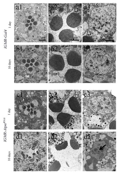
Figure 6.
EM analyses revealed that ATPα knockdown photoreceptors in 10-day-old flies underwent neurodegeneration as characterized by apoptotic/hybrid cell death. Those photoreceptors lost rhabdomeres, and the remaining rhabdomeres were reduced in size (d). Apoptotic features, including condensed nuclei and dark chromatin clumps (indicated by arrow, d3), accompanied by necrotic changes such as cytoplasmic edema manifested by vacuolation (indicated by asterisks, d2 and d3) were observed in the cell bodies of degenerating photoreceptors. Although rhabdomeres in 1-day-old knockdown flies appeared to be slightly smaller than those of control lGMR-Gal4 flies, no obvious degeneration was observed (c2 and c3).

