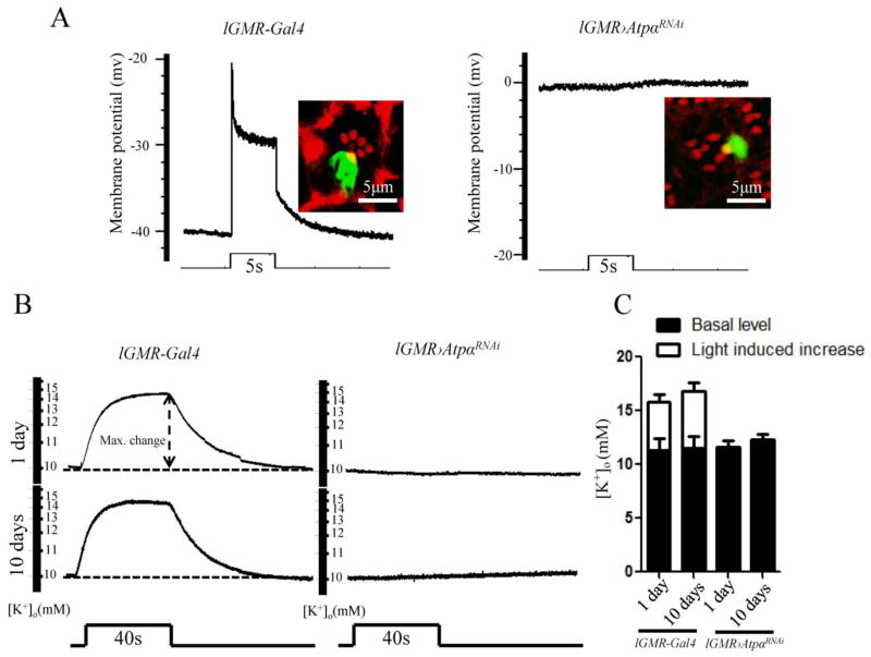
Figure 7.
The photoreceptors in ATPα knockdown flies lost their ability to maintain the K+ gradient. A, Intracellular recordings of photoreceptors were performed in 1-day-old control lGMR-Gal4 and ATPα knockdown flies. The resting membrane potential of the photoreceptor in the dark is approximately −40 mV in controls (n=10) and 0 mV in knockdown flies (n=10). Light stimulation failed to trigger any electrical response in the ATPα knockdown photoreceptors. To confirm the cellular identities, photoreceptors were electrically injected with neurobiotin, and the labeled cells were visualized by streptavidin-Alexa Fluor 488 conjugate and rhodamine phalloidin. B, Retinal [K+]o levels were measured with double-barrel K+-selective microelectrodes. The [K+]o level was increased by a 40-sec light stimulation in 1-day-old and 10-day-old control flies but not in ATPα knockdown flies. Dashed line: signal level in the dark. C, Comparison of basal [K+]o levels and the 40-sec light-stimulated [K+]o levels shows an increase in the retina between the control and ATPα knockdown flies. Asterisk (*): p< 0.01; Two-tailed t-test. Error bars denote S.E.M.

