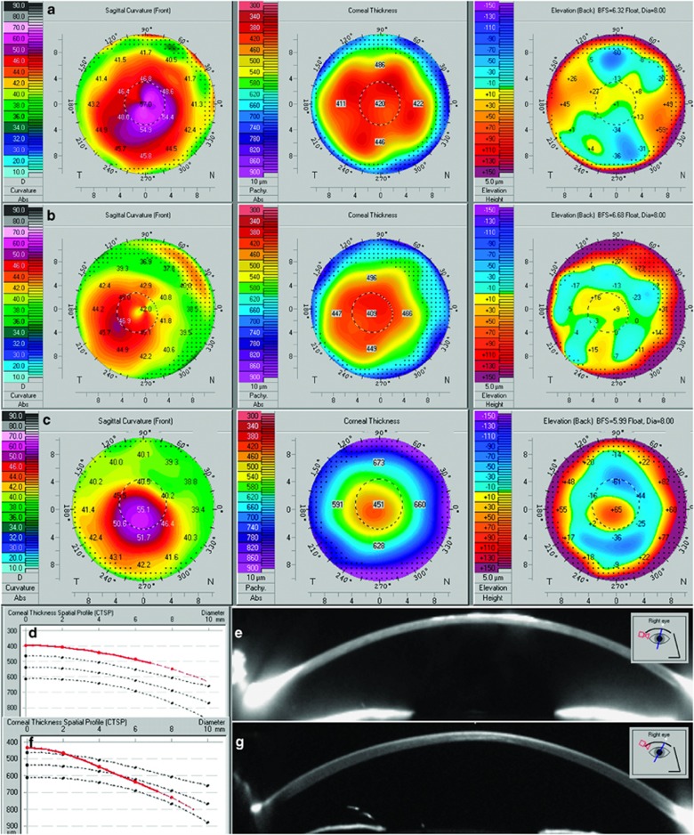
Figure 1.
Scheimpflug imaging of macular corneal dystrophy and keratoconus corneas. Anterior sagittal curvature, pachymetry, and posterior elevation maps of the right cornea of proband 1 (a) and the right cornea of proband 5 (b), note diffuse thinning on pachymetry map and lack of posterior elevation in both a and b. Right cornea of a patient with keratoconus, note localized ectasia observed on all three maps (c). Corneal thickness spatial profile of the right cornea of proband 5, note uniformly thin cornea shown by a red line (d). Single Scheimpflug image of the right cornea of proband 1 further documenting diffuse thinning; imbedded image shows meridian of capture (e). Corneal thickness spatial profile of keratoconic cornea, note progressive thinning towards the corneal center (f). Single Scheimpflug image of the right cornea of a keratoconus patient documenting localized paracentral thinning, imbedded image shows meridian of capture (g).

