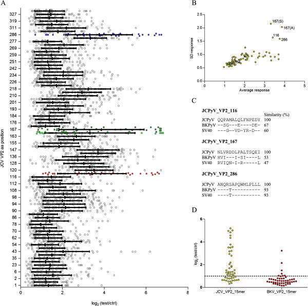
Figure 1.

Identification of JCPyV_VP2_167-15mer as immunoreactive peptide. (A) Distribution of the JCPyV VP2 polyomavirus 15-mer peptide microarray signals obtained with plasma samples from 49 HSs, diluted at 1:200. Peptides with the largest signal distribution are indicated in red (JCPyV_VP2_116-15mer), green (JCPyV_VP2_167-15mer, light green is S175 variant, dark green is A175 variant) and blue (JCPyV_VP2_286-15mer). (B) Average signal in microarray vs. standard deviation of signal over the different subjects, plotted per peptide. (C) Similarity between peptides specific to JCPyV, BKPyV and SV40 for the 3 selected JCPyV peptides. (D) Plasma antibody reactivity of 50 HSs against JCPyV_VP2_167-15mer and BKPyV_VP2_167-15mer, with data presented as log2 (signal of test sample / signal of the no-sample control). Samples were diluted at 1:200 and detected in a peptide ELISA.
