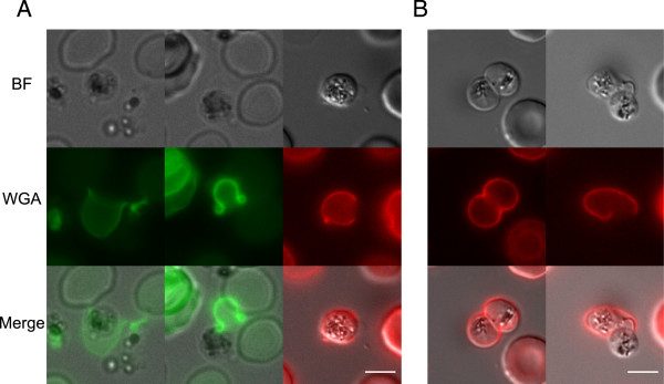
Figure 3.

Red blood cell membrane disruption in gametes egress. A) Gamete egress observed on live (left and central panels, WGA-Alexa488 staining) and fixed (right panel, WGA-Texas Red staining) parasites. B) Two gametes within the same red blood cell. The host cell shows in one case an intact membrane (left panel) and a single opening at the start of gamete egress (right panel). Scale bars: 5 μm. BF: bright field. WGA: wheat germ agglutinin.
