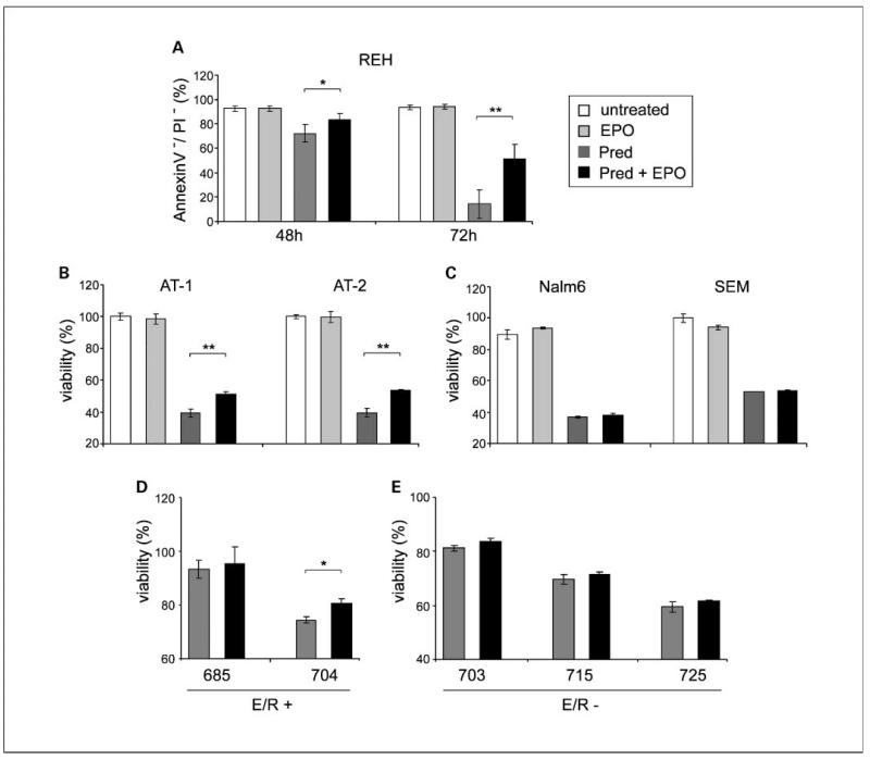
Fig. 4.
EPO attenuates prednisone-induced apoptosis in ETV6/RUNX1-positive leukemias. A, apoptosis rates were determined byAnnexinV/propidium iodide staining in REH cells cultured for 48 and 72 h in the presence of prednisone (Pred; 1mg/mL) and EPO (50 units/mL). The percentage of viable, AnnexinV-positive/propidium iodide-positive cells is depicted. Mean ± SD of four experiments. Evasion from prednisone-induced apoptosis by EPO in AT-1and AT-2 cell lines (B), Nalm6 and SEM (C), and ETV6/RUNX1-positive and ETV6/RUNX1-negative primary leukemias (D and E). Cells were exposed to prednisone (50 μmol/L) in the presence and absence of EPO (50 units/mL) and viability was assessed by 3-(4,5-dimethylthiazol-2-yl)-2,5-diphenyltetrazolium bromide assay after 72 h. The increase in cell viability by EPO is indicated in percentage. Mean ± SD from triplicates (in cell lines, one of three independent experiments, in primary leukemic cells from one experiment). *, P < 0.05;**, P < 0.01.

