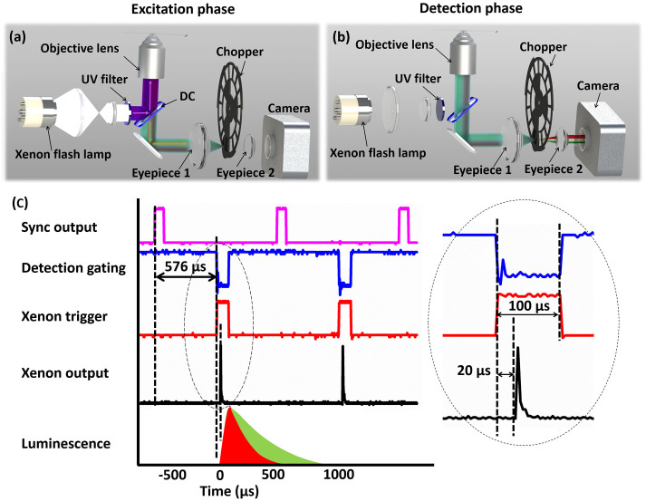
Figure 1. Schematic diagrams of the multi-colour TGL microscope.
(a) In the excitation phase, a pulsed excitation light illuminates the sample, while the chopper stops the luminescence/autofluorescence being captured by camera. (b) In the detection phase, the excitation is turned off, and the chopper allows the luminescence to reach the camera. (c) The time sequence of the system is shown, with every repetition cycle containing a gating window of 88 μs and a detection window of 968 μs. Each flash pulse is released 20 μs after the trigger and last around 17 μs.

