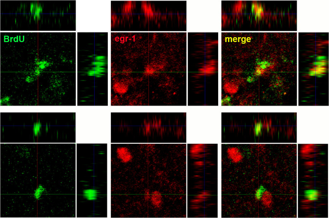
Figure 11. Two examples of confocal images showing newborn cells (BrdU+) 1 month post lesion that expressed singing-induced egr-1 in the recovered part of LArea X.
To confirm that the cells are double labeled, three planes of the confocal image are shown: dorsal y view (largest panel), lateral x view (upper panels), and lateral z view (side panels). Crossing of the perpendicular lines are where the centers reference for each panel.

