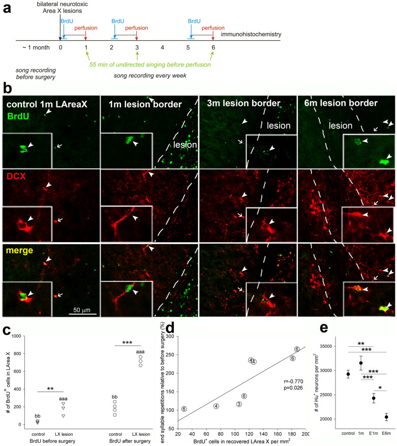
Figure 9. Timeline and Area X tissue recovery.
(a), Timeline. In different groups of birds, songs were recorded from about 1 month before and up to 6 months after the surgery. The cell division marker BrdU was injected for 7 days at 0, 2, or 5 months after the surgery and birds sacrificed 30 days after the first injection, after performing 55 min of undirected singing. (b), Representative photomicrographs of control LArea X and lesion border 1, 3 and 6 months after the neurotoxic injury showing higher numbers of new (BrdU+) young neurons (DCX+) with increasing age. The dashed lines highlight the visible boundary between the lesion and nonlesioned or recovering parts of LArea X. Arrows and arrowheads point to double labeled DCX+/BrdU+ neurons. Inserts show double-labeled neurons magnified 6x relative to the main panels. Note the high number of BrdU+ cells inside the lesion at 1 month and higher number of DCX+ neurons inside the lesion at 3 and 6 months in comparison to 1 month. The DCX+ neurons at 1 and 3 months are rounded and with few dendritic processes while at 6 months their expression was less and the cells appeared to have more dendritic processes. (c), New cells (BrdU+) incorporated in the recovered part of LArea X with BrdU injected during 7 consecutive days either before or immediately after the surgery. ** p < 0.01, *** p < 0.001, comparison between control and lesioned birds; ‘aaa’ p < 0.001 for the comparison between lesioned birds with BrdU injections before and after surgery; ‘bb’ p < 0.01 for the comparison between controls (ANOVA followed by Fisher’s PLSD post hoc test). (d), Correlation of the number of new (BrdU+) cells in the recovered part of LArea X and number of end syllable repetitions after neurotoxic lesion (linear regression). The numbers in circles indicate survival of the bird (in months). (e), Neuronal density in MSt adjacent to the lesion 1 month after neurotoxic lesion (1m) and 1 and 6 months after electrolytic lesion (E1m and E6m). * p < 0.05, ** p < 0.01, *** p < 0.001 (ANOVA followed by Fisher’s PLSD post hoc test).

