
Figure 2.

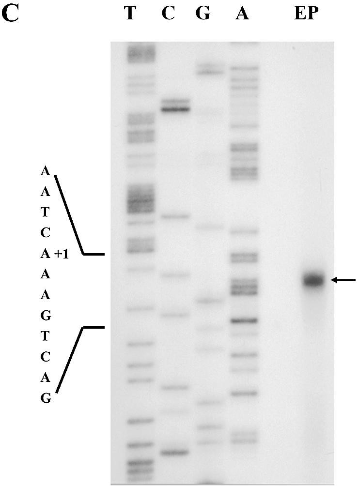
Identification of the transcriptional start site. (A) Schematic representation of the 5′-flanking region of dsps2, the open reading frame (ORF), the 300 bp probe, the 161 bp protected fragment and the primer used for sequencing. (B) Autoradiogram of RPA: lane 1, the probe used for RPA; lane 2, the protected probe from RNaseA/T1 by Drosophila RNA; lane 3, the negative control using yeast RNA. The assay was performed as described in Materials and Methods. The sequence of the 5′-flanking region of dsps2 that was generated by using the RPA primer is shown on the left. (C) Results of primer extension. The arrow designates the extended band in the lane marked as EP (Extended Product). The sequencing reaction surrounding the transcription start site is shown on the left.