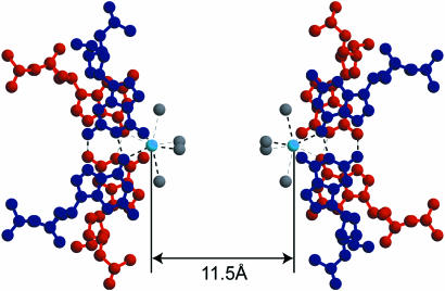
Figure 5.
Two potassium cations bound to four guanine bases in the central parts of the split quadruplexes. Four water molecules cover the outer side of each cation. The G-duets are colored in red or blue. The value indicates the K…K distance in G1-I-highK. Light blue and gray spheres represent potassium cations and water oxygen atoms, respectively. Broken lines indicate eight coordination bonds around the respective potassium cations and possible hydrogen bonds.

