Figure 1.
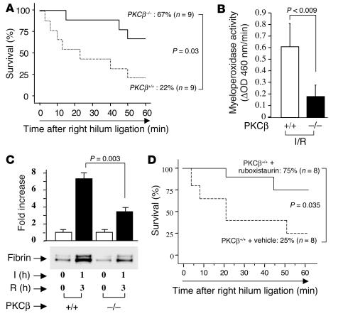
Murine model of lung ischemia/reperfusion (I/R): effect of PKCβ. (A and D) Survival analysis. PKCβ+/+ and PKCβ–/– (A) and PKCβ+/+ mice fed vehicle chow or ruboxistaurin chow (D), respectively, were subjected to left-lung ischemia for 1 hour and reperfusion for 3 hours. Blood flow to the uninstrumented right lung was then blocked, and mortality was determined after 1 hour with only the left lung in the circulation. (B) Myeloperoxidase activity. After I/R, lung samples were harvested from PKCβ+/+ and PKCβ–/– mice and subjected to myeloperoxidase activity assay (n = 5). (C) After left-lung I/R as described above, animals received systemic heparin and were sacrificed. Lung protein extract was digested with plasmin and subjected to SDS-PAGE (7.5%; 0.2 ∝g of total protein/lane). Immunoblotting with anti-fibrin antibody was performed. Data are shown as mean ± SEM of five experiments.

