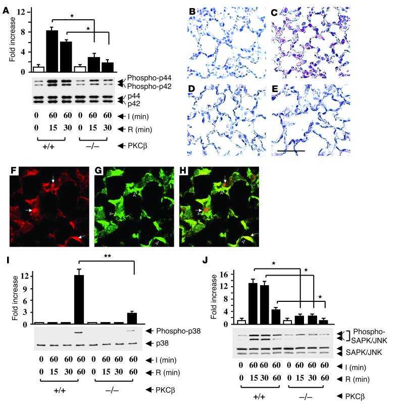
Figure 3.
Ischemia/reperfusion-mediated activation of MAPKs in the lung. PKCβ+/+ and PKCβ–/– mice underwent the indicated period of left-lung I/R. Animals were sacrificed and protein extracts from the I/R and uninstrumented lung were prepared and subjected to SDS-PAGE (12%, 50 ∝g of protein/lane). Immunoblotting with phospho-p44/42 MAPK antibody or total p44/42 MAPK antibody (B), phospho-p38 MAPK antibody, or total p38 MAPK antibody (I), and phospho-SAPK/JNK antibody or total SAPK/JNK antibody (J) was performed. Data are shown as mean ± SEM of five experiments. Immunohistochemical analysis of phospho-p44/42 expression in murine lung from uninstrumented (B) or I/R (C) PKCβ+/+ mice, and from uninstrumented (D) or I/R (E) PKCβ–/– mice was performed. Scale bar: 50 ∝m. I/R lungs from PKCβ+/+ mice were subjected to immunofluorescence microscopy and double stained with an anti–phospho-p44/42 antibody (red) (F) and an anti-macrophage antibody (F4/80, green) (G). The merging of F (pERK1/2) and G (F4/80) is shown in H. Arrows in F and arrowheads in G indicate dually stained cells Original magnification in F–H, ∞1,000. *P < 0.001; **P = 0.0041.

