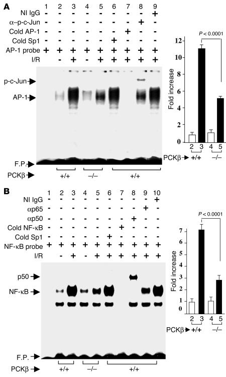
Figure 6.
I/R-mediated induction of AP-1 and NF-κB in the lung: effect of PKCβ. Mice underwent left-lung I/R or no instrumentation. Mice were sacrificed, and nuclear extracts were prepared from the lung and subjected to EMSA with 32P-labeled consensus AP-1 (A) and NF-κB (B) probes. Where indicated, nuclear extracts from uninstrumented (lane 2 and lane 4) and I/R (lane 3 and lane 5) lung were incubated with 32P-labeled AP-1 (A) or NF-κB (B) probe alone. Nuclear extracts from I/R lungs of PKCβ+/+ mice were incubated with 32P-labeled AP-1 (A) or NF-κB (B) probe in the presence of either a 100-fold molar excess of Sp1 (cold Sp1; lane 6 in A and B), AP-1 (cold AP-1; lane 7 in A), or NF-κB (cold NF-κB; lane 7 in B), and either anti–p-c-Jun IgG (lane 8 in A), anti-p50 IgG (lane 8 in B), and anti-p65 IgG (lane 9 in B) or nonimmune (NI) IgG (lane 9 in A and lane 10 in B, respectively). F.P., free probe.

