Figure 3.
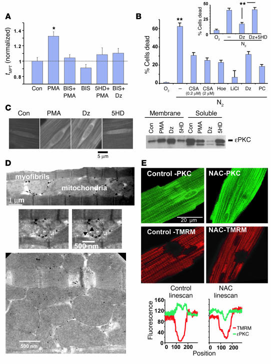
Mechanisms of protection. (A) MPT susceptibility to ROS (tMPT) can be regulated by the mitoKATP: role of PKC. *P < 0.001 vs. control (Con). (B) Activation of distinct protection pathways improves cell survival to a similar degree in the hypoxia/reoxygenation protocol used to assess tMPT. Hypoxia/reoxygenation groups included treatment with cyclosporin A, Hoe, Li+, Dz, and PC. The protective effect of Dz is abolished by 5HD (inset). **P < 0.02. (C) Translocation of εPKC toward mitochondria, induced by mitoKATP activation. The panels on the left represent immunostained cardiac myocytes (15 ∞ 15 ∝m2 region surrounding the nucleus, shown for technical consistency). The immunoblot on the right shows that both Dz and PMA induce εPKC translocation from the soluble to the membranous cellular fraction. (D) Transmission electron microscopy of immunogold-labeled εPKC in a cardiac myocyte from a heart treated with PMA (100 nM, 15 minutes), demonstrating mitochondrial membrane localization (right middle panel; dashed circle outlines a mitochondrial profile); immunolabeling is absent in control (not shown). (E) ROS-induced PKC translocation toward mitochondria. Photoexcitation-mediated MPT induction in an approximately 10 ∞ 10 ∝m2 region in TMRM-loaded cardiac myocytes (middle panels, red), and εPKC immunostaining (top panels, green) in the same cells. The right panels show effects of the ROS scavenger NAC. The bottom panels compare the εPKC labeling through the photoexcited regions.

