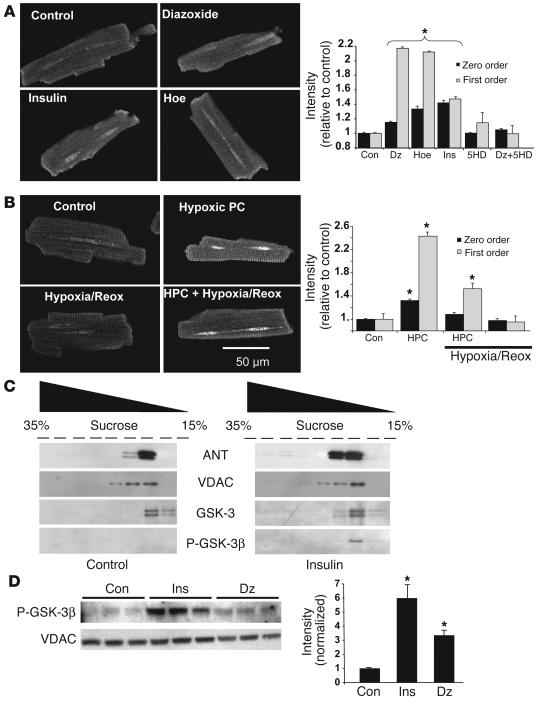
Figure 8.
Localization and regulation of a mitochondrial GSK-3β pool during protection signaling. (A and B) P-GSK-3β immunocytochemical labeling: changes in average versus compartmentalized signal intensity determined from 2D Fourier analysis (see text; images of cells exposed to 5HD and to Dz plus 5HD not shown). *P < 0.01 (all bars under brace) vs. respective control. (C) Immunoblots of mitochondrial membrane sucrose gradient fractions, from control and insulin-treated (30 nM) rat hearts, probed with adenine nucleotide translocator (ANT), voltage dependent anion channel (VDAC), GSK-3, and P-GSK-3β. (D) Immunoblot of total mitochondrial proteins isolated from control, insulin-treated (30 nM), and Dz-treated (30 ∝M) rat hearts. **P < 0.03 vs. control.

